Neuro Ophthalmology
What is Neuro-Ophthalmology?
Neuro-Ophthalmology is an exclusive subspecialty of Ophthalmology, focused on the evaluation and diagnosis of neurological conditions that affect the visual system.
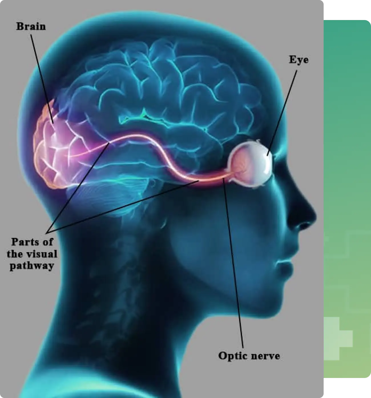
The optic nerve, which extends from behind the eyeball, is responsible for transmitting visual information to the brain. Because of this vital connection, Neuro-Ophthalmology addresses a wide range of conditions that simultaneously impact both the eye and the brain.
This field bridges the gap between neurology and ophthalmology, offering expert care for complex disorders where vision and the nervous system intersect.
Visual loss can result from a variety of causes — including acquired or inherited optic nerve disorders, or conditions that affect intracranial visual pathways due to stroke or brain tumours. In some cases, the cause of vision loss may remain unexplained, requiring a detailed and in-depth diagnostic evaluation.
We also manage conditions involving double vision caused by abnormal eye movements, which often require careful and coordinated care.
Many of our patients present with complex neurological or systemic conditions that require co-management across specialties such as Neurology, Neurosurgery, and Neuroradiology.
Comprehensive Neuro-Ophthalmic Care
At Narayana Nethralaya, we believe in a holistic approach to patient care. Our goal is to offer the most effective, tailor-made treatment plans, thoughtfully weighing the risks and benefits of all available options.
Our dedicated team includes experienced Neuro-Ophthalmologists, and a Neurologist (who is available twice a week at our NN1 branch to ensure coordinated, multidisciplinary care.)
Symptoms of Neuro-Ophthalmic Conditions

Blurred Vision
One of the most common symptoms seen in neuro-ophthalmic conditions is blurred vision. This may affect one or both eyes and is often:
- Sudden in onset
- Sometimes accompanied by pain
- Caused by optic nerve-related disorders
For these conditions, prompt medical evaluation is crucial.

Double Vision
Do you suddenly see two objects instead of one?
This condition, known as double vision or diplopia, may have a neurological or neuromuscular cause and should not be ignored.
Droopy Eye Lids (Ptosis)
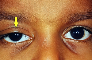
Noticing a sudden drooping of one or both eyelids?
This condition, medically known as ptosis, can occur with or without double vision and may indicate a neurological or neuromuscular disorder.
Eye Motility Disorders
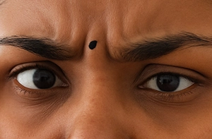
Do your eyes seem out of sync?
Is one eye not moving as freely as the other?
This may be a sign of an eye motility disorder, which can arise from:
- Eye muscle weakness
- Or other neurological conditions
These disorders often result in intermittent or constant double vision (diplopia).

Headache
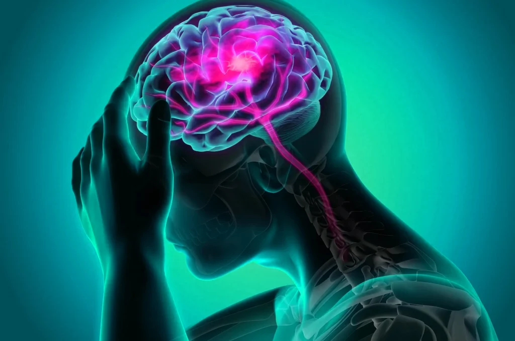
Headaches can sometimes be more than just pain — they may be a sign of an underlying neurological condition, especially when accompanied by:
- Visual blurring
- Buzzing in the ears (tinnitus)
- Nausea or vomiting
These symptoms may indicate raised intracranial pressure, which requires immediate medical attention.
Other types, like migraine headaches, may or may not involve visual disturbances and are often triggered by specific factors. Migraines are frequently accompanied by:
- Sensitivity to light (photophobia)
- Sensitivity to sound (phonophobia)
A sudden, severe headache, especially when combined with fever, weakness, numbness, or other neurological symptoms, needs immediate attention and management.

Facial Paralysis
Noticing a sudden inability to close one eye?
Are the muscles of facial expression on one side of your face not moving?
These could be signs of facial paralysis
Optic neuropathies:
Diseases affecting the optic nerve like optic neuritis, ischemic optic neuropathy, autoimmune optic neuropathy, compressive optic neuropathy, nutritional optic neuropathy, toxic optic neuropathy.
Ocular motility disorders:
characterized by partial of complete inability to move either one or both the eyes due to central and peripheral nervous system diseases like myasthenia gravis and ocular motor cranial nerve palsies following trauma (injury), tumours, ischemia due to systemic diseases like hypertension or diabetes mellitus.
Demyelination disorders
affecting the visual pathway formed by the optic nerve, optic chiasma, optic tract and optic radiations.
Tumours
like pituitary adenoma, craniopharyngioma, meningioma causing compressive optic neuropathy which cause a direct pressure on the optic nerve and other tumours of the brain affecting the visual pathway and cause of raised intracranial pressure.
Other conditions causing raised intracranial pressure
such as idiopathic intracranial hypertension, and cerebral venous thrombosis, which can result in permanent and profound loss of vision in the long term
Cerebrovascular disorders
with visual loss, transient ischemic attacks with transient visual loss
Optic Neuropathies
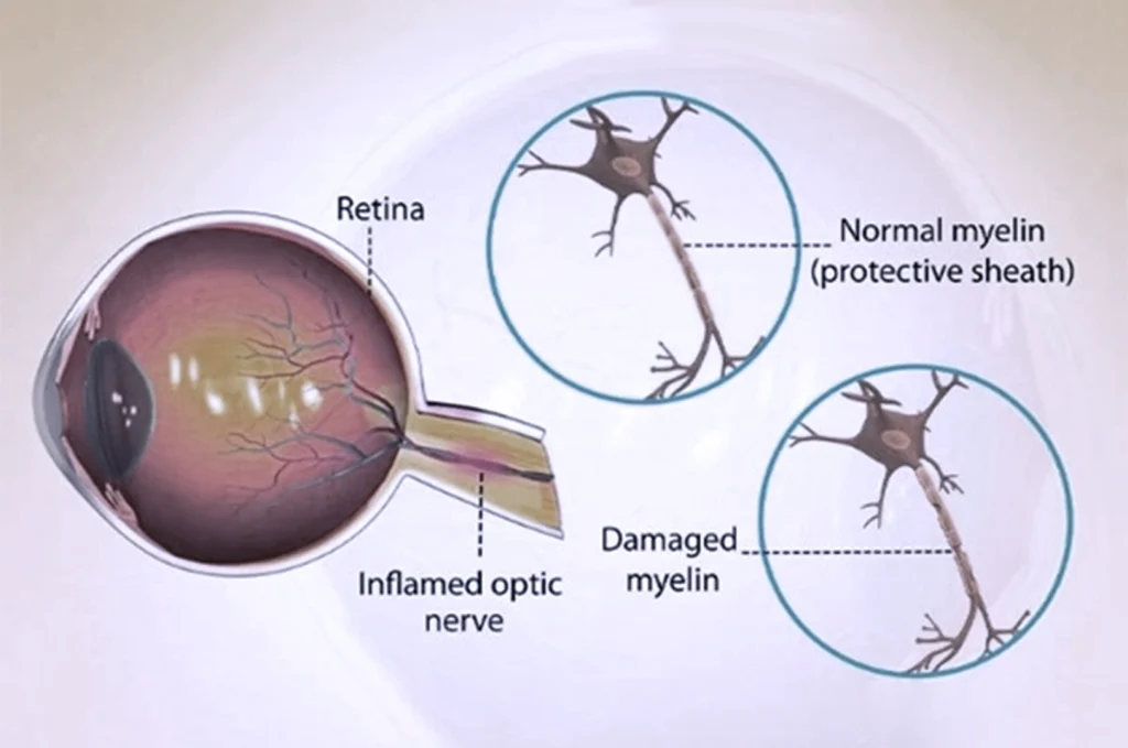
Optic neuropathies refer to a group of conditions that affect the optic nerve, which carries visual information from the eye to the brain. Damage to this nerve can lead to sudden or progressive vision loss. It is often associated sudden visual blurring, reduced brightness/contrast and impaired colour vision. It is commonly seen in inflammation and autoimmune conditions often requiring prompt evaluation.
Common types of optic neuropathies include:
- Optic neuritis – often associated with inflammation and autoimmune conditions
- Ischemic optic neuropathy – due to reduced blood flow to the optic nerve
- Autoimmune optic neuropathy – linked to disorders like lupus or sarcoidosis
- Compressive optic neuropathy – caused by tumours or masses pressing on the optic nerve
Nutritional or toxic optic neuropathy – due to vitamin deficiencies or toxic exposures (e.g., alcohol, medications)
Conditions Causing Raised Intracranial Pressure
Certain neurological conditions can lead to raised pressure inside the skull (intracranial pressure), affecting both brain function and vision.
Two common conditions include:
- Idiopathic Intracranial Hypertension (IIH)
- Cerebral Venous Thrombosis (CVT)
These conditions often present with
- Severe persistent Headaches
- Visual blurring
If not detected and treated early, they can result in permanent and profound vision loss.
Eye Movement Disorders
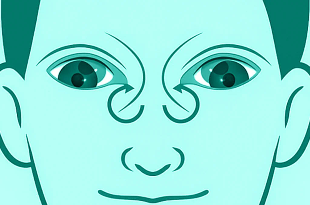
Eye movement disorders refer to the partial or complete inability to move one or both eyes, often resulting from abnormalities in the central or peripheral nervous system.
Common causes include:
- Cranial nerve palsies due to ischemia (often from systemic conditions like diabetes or hypertension)
- Trauma-related nerve injury
- Brain tumours or compressive lesions
- Neuromuscular junction disorders such as myasthenia gravis
Intracranial Tumors
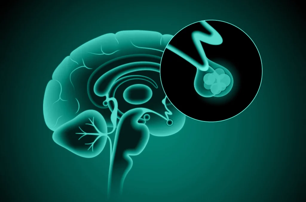
Certain brain tumours can lead to visual disturbances either by:
- Direct compression of the optic nerve or optic pathways, or
- Indirectly, through raised intracranial pressure
Cerebrovascular Disorders
Cerebrovascular disorders, such as strokes or transient ischemic attacks (TIAs), can lead to visual impairment when the visual cortex in the brain is affected.
This may result in:
- Partial or complete vision loss
- Loss of vision in one side or part of the visual field (visual field defects)
- Symptoms that may be transient or permanent, depending on the severity and location of the vascular event

Conditions That Cause Abnormal or Involuntary Movements of the Face or the Eye
One might experience twitching or involuntary movements of the facial muscles.
Involuntary movement of the eyeballs called nystagmus may be congenital or acquired (due to a central or peripheral nervous system cause)
Asymmetric pupil size
Also called anisocoria , can be due to abnormalities in Pupillary spinchter muscles or may be diagnostic clues to underlying neurological pathologies.
Diagnostics
At Narayana Nethralaya, we are equipped with advanced and state-of-the-art electrodiagnostic testing facilities to accurately assess the function and health of the optic nerve and the visual pathways.
Our diagnostic capabilities include:
Humphrey Visual Field Analysis
Used to evaluate visual field defects and monitor conditions like glaucoma, optic neuropathies, and neurological vision loss.
Optical Coherence Tomography (OCT)
A high-resolution imaging tool that provides cross-sectional views of the retina and optic nerve, essential for diagnosing and monitoring optic nerve and retinal disorders.
B-Scan Ultrasound
Useful in visualizing the posterior segment of the eye, especially in cases where media opacities prevent direct viewing.
Electrophysiology
Includes specialized tests such as:
- VEP (Visual Evoked Potential) – to assess the function of the visual pathway from the eye to the brain
- ERG (Electroretinogram) – to evaluate retinal function
- EOG (Electrooculogram) – to assess the function of the retinal pigment epithelium.
These cutting-edge tools allow our neuro-ophthalmologists to make accurate diagnoses and tailor effective treatment plans for each patient.
Patient Process Flow
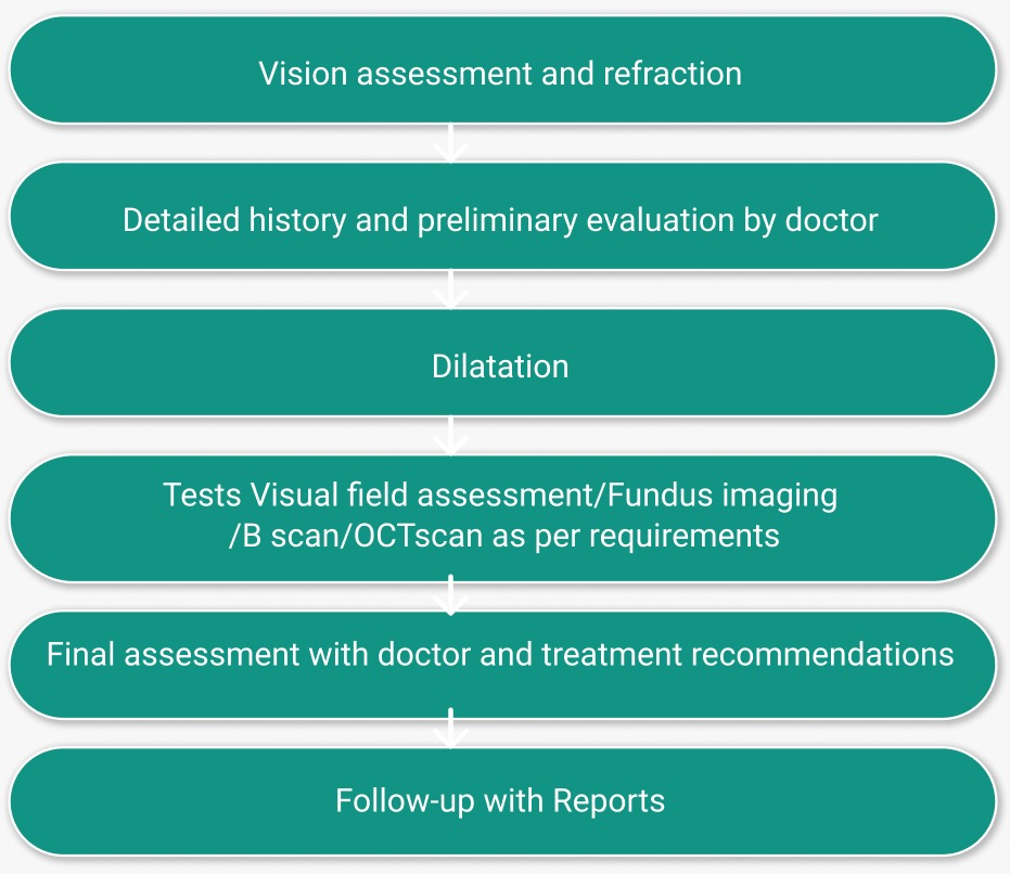
If you have constant blurred vision, double vision, sudden onset of drooping of eyelids, headaches with vomiting & blurring of vision, facial paralysis or abnormal involuntary movements of the eyes and face, you should schedule an appointment with an ophthalmologist to get your eyes checked immediately. Your initial consultation with the doctor will approximately take 2 hours if you do not require cross-consultation and up to 4 hours if you require cross-consultation. During your consultation, our doctors and counsellors will determine the best course of action for your visual needs, go over the risks and benefits of treatment, and help you choose the best procedure that is suitable for preserving/improving your vision. We suggest you bring a family member or friend with you to help you with your decision-making.
Now Eye Know Videos
From Headache to Double Vision: 5 Common Neuro-Ophthalmology Symptoms Explained | #DoctorSpeak
Frequently Asked Questions
When should I consult a neuro ophthalmologist?
You must consult a neuro ophthalmologist when there is a sudden loss of vision, sudden onset of visual field defects, when one has double vision, chronic progressive visual loss, drooping of eyelids, transient visual loss, unequal pupil size. just to mention a few.
What does a neuro ophthalmological examination include?
It includes assessment of visual acuity, color vision, pupillary reaction, assessment of the optic nerve appearance and function, assessment of the visual pathway from the eye to the visual cortex in the brain, assessment of extraocular movements, assessment of the field of vision and other tests as and when required.
What special tests are required in visual loss /blurring?
Any of these tests may be required in isolation or in combination.
- Automated Visual fields
- VEP
- OCT
- Carotid and Vertebral Doppler
- CT brain and orbit
- MRI brain and orbit
- MRA brain and neck vessels
- MRV brain
- CT angiogram of brain and neck vessels
- Blood tests when necessary
What is the optic nerve?
The optic nerve is a bundle of nerve fibers which conduct visual information from the eyes to the visual cortex of the brain.
What is optic neuritis?
Optic neuritis is an inflammation of the optic nerve. The patient experiences sudden blurring of vision in one or both eyes with pain in the eye/eyes, painful eye movements, color vision abnormalities, and sometimes flashing lights.
What are the common causes of optic neuritis?
The most common causes are demyelinating illnesses like multiple sclerosis and neuromyelitis optica, viral demyelination. Other causes include infectious diseases, autoimmune diseases.
Do patients with optic neuritis recover vision?
Most certainly, they do so, but they must consult the neuro ophthalmologist and neurologist immediately. The sooner the treatment is started, the better the outcome.
Once the optic nerve is damaged, can it recover?
Unfortunately, damage to the optic nerve fibers is irreversible. They cannot regenerate on their own.
Where is the vision center in the brain?
The vision fibers in the optic nerves form the optic tracts, the optic radiations and finally end in the visual cortex in the occipital lobe at the back of the brain.
Can double vision be treated?
Yes, it can be treated. Most recover within a month or two. Sometimes double vision due to trauma/accidents may not recover fully and may require special prisms or surgical correction.
What are the common causes of headache in neuro ophthalmology?
Headache may occur in migraine, vascular headache, tension headache, cluster headache, meningitis, raised intracranial pressure due to any cause, double vision, inflammation of the eye which may include optic neuritis, inflammation behind the eye in conditions like para nasal sinusitis, orbital apex syndrome, Tolosa Hunt syndrome, cavernous sinus syndrome.
What are the common causes of raised intracranial pressure?
The common causes are idiopathic intracranial hypertension, cerebral venous thrombosis, intracranial tumors, meningitis, intracranial hemorrhage, traumatic brain injury.
What does the patient experience when there is raised intracranial pressure?
The common symptoms of raised intracranial pressure include severe headache with nausea, projectile vomiting, transient visual blurring, tinnitus, double vision, loss of consciousness.
Can stroke cause loss of vision?
Yes it can. If the clot or bleed in the brain involves the optic radiation or the visual cortex there will be visual loss along with other features.
Can a stroke cause double vision?
Yes, it can. If the stroke involves the brain stem, where the nerves controlling eye movements originate, the patient can have sudden onset of double vision.
Why does somebody have double vision?
Double vision may be monocular or binocular. Monocular double vision is not neurological. Binocular double vision is usually neurological in origin. When any nerve which controls eye movements is affected, double vision can occur.
Can smoking and alcohol affect vision?
Yes, they can affect your vision directly or indirectly.
Patient Testimonials
Prism Glasses To Fix Vision Problems After Brain Injury | Patient Testimonial
A 13 year old girl came with sudden blurring of vision in the right eye, with pain since 1 week and right sided headache.
On evaluation her vision was very poor in the right eye. Though she did not complain of visual disturbances in the left eye, detailed evaluation showed that the left eye was also affected, but to a lesser extent. She was diagnosed to have optic neuritis in both eyes. Neuroimaging showed optic neuritis in both eyes and additional changes in the brain suggestive of neuromyelitis optica spectrum disorder.
She was referred to our Consultant Neurologist who immediately admitted and treated her. She recovered her vision completely in both eyes after treatment and was on subsequent treatment with disease modifying drugs. The field defects in both eyes also had recovered completely. On follow up she was doing well and responding extremely well to treatment. She needed long term treatment and regular follow-up. The parents were explained regarding the condition and the need for regular eye check up and also regular followup with the Neurologist.
The parents and the child were very happy that the vision had recovered completely in both eyes.
A 27 year old gentleman came to Narayana Nethralaya with poor vision in the right eye which was progressively worsening since 6 months. He also had headache since 4 years which was worse since 6 months.
He underwent a thorough ophthalmic evaluation. His vision in the right eye was very poor and both optic nerves appeared pale or had optic disc pallor. He also had poor vision in the outer half of the field of vision in both eyes which was confirmed after evaluation. A compressive mass lesion at the optic chiasm was suspected. Neuroimaging confirmed this and he was diagnosed as having a Rathke’s cleft cyst. He was immediately referred to our Consultant Neurosurgeon, who evaluated him and the gentleman underwent surgery for removal of the tumor.
2 months later , on followup, his vision in both eyes had recovered to normal. His field defects in both eyes recovered completely.
The patient had no visual symptoms and no headache. On subsequent follow up, he was doing well after 2 years also and the follow up neuroimaging showed no recurrence of the tumor. The patient was extremely happy with the results.
Our patient care philosophy
At Narayana Nethralaya, “Quality of Care” and “Patient Safety” is our priority. Concern for our patients’ well being is at the core of what we do, and what drives us. Five units of Narayana Nethralaya are NABH Accredited – the highest national recognition for quality in patient care and safety. Our Neuro ophthalmology team help patients make an informed treatment choice on the type of treatment and surgery that is best suited for their lifestyle. We have an exclusive counseling team to address any doubts or questions that people may have about Neuro ophthalmology treatment options and procedures.

