The eye is comparable to a camera with a lens in front and a film (which we call retina) behind. Retina is one of the 4 important structures which is responsible for good quality and quantity vision which is placed approximately 2 centimetres behind the cornea. The light entering the eye reaches the retina and is further relayed via the optic nerve to the brain. Thus, severe vision loss can occur in diseases affecting the vitreous, retina and optic nerve. The modern methods of diagnosis and treatment has made it possible to manage these disorders satisfactorily.
Narayana Nethralaya has a fully-functional retinal department conducting retinal surgeries and medical management of retinal disorders in all its 5 branches. The retinal specialist team is the largest in the entire state of Karnataka and is one of the major referral center which deals with the management of diabetic retinopathy, retinal detachment, ARMD, infections, trauma & various other retinal disorders. Our retinal specialist team comprising of clinical specialists and basic scientists have pioneered the clinical care, research efforts, and development of all major innovations in retina care for the last 39 years. These highly-skilled and experienced surgeons have been intimately involved in the development of most of the treatments for many retinal diseases, including the latest treatments for macular degeneration and diabetic retinopathy.

Retinal imaging is to take pictures of your retina i.e, back of your eye. Imaging is done using different techniques as per the diagnosis. It helps your doctor to detect the earlier/subtle changes of retinal diseases which cannot be identified on routine eye examination. Early detection of diseases helps to treat the disease properly.
Diabetic retinopathy occurs secondary to deterioration of blood vessels in the retina as a complication of diabetes mellitus. The Breakdown of retinal blood vessels may result in fluid leaking into the center of the retina (macular edema) or abnormal blood vessels formation on the surface of the retina (neovascularization) which can bleed and scar resulting in loss of central and possibly peripheral vision.
Age-related macular degeneration (AMD) is a common, complex, degenerative condition affecting people aged 50 years and above causing central vision loss, which results in difficulty to read, drive, or see someone’s face on progression to more advanced stages.
Retinal vein occlusion is a blockage in the ocular blood vessel that can result in vision loss. The retinal veins carry away used blood from the retina and when one of these veins are blocked, the used blood cannot drain away which causes swelling and hemorrhage (bleeding).
Age-related macular degeneration (AMD) is a common, complex, degenerative condition affecting people aged 50 years and above causing central vision loss, which results in difficulty to read, drive, or see someone’s face on progression to more advanced stages.


Narayana Nethralaya has fully equipped state of art vitreo retinal surgical suites in each of its branches. The surgeries are performed for a variety of retinal disorders like retinal detachment, advanced diabetic eye disease, vitreous hemorrhage, endophthalmitis, macular hole and epiretinal membrane using the latest small gauge micro incisional vitrectomy system (MIVS). The institute also boasts of having the 3D Ngenuinity visualisation system which allows the surgeon to perform a heads-up vitreoretinal surgery.
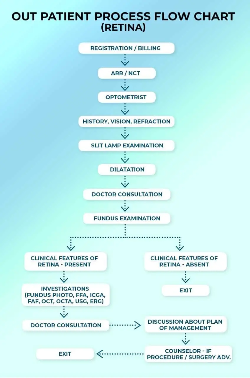
If you have sudden appearance of floaters, blurring of vision, flashes of light in one or both eyes, loss of vision or a “shadow” over the visual field, you should schedule an appointment with an ophthalmologist to get your eyes checked immediately. Your initial consultation with the doctor will approximately take 2 hours if you do not require cross-consultation and up to 4 hours if you require cross-consultation. During your consultation, our doctors and counsellors will determine the best course of action for your visual needs, go over the risks and benefits of treatment, and help you choose the best procedure that is suitable for preserving and improving your vision. We suggest you bring a family member or friend with you to help you with your decision-making.
Retina is the photosensitive inner lining of the eye (similar to the photographic film of a camera). When light enters the eye, the image is first formed on the retina and then sent to the brain via the Optic Nerve
Macula is the centre of the retina. For practical purposes, all fine vision is located at the macula.
It is recommended that people with high numbers, all diabetics and people > 40 years of age get the retina checked at least once a year.
By putting some drops called “mydriatics” which dilate the pupils. The retina is the examined by an instrument called the “indirect ophthalmoscope”.
Will someone need to accompany me for the check-up? – the pupillary dilatation can often cause significant blurring of vision for 3-4 hours. It is therefore recommended not to drive after the test. It is for the same reason that we often ask someone to accompany the patient for the test. (especially patients with already poor vision)
Diabetes affects the retinal capillaries and other blood vessels. The effects of these on the retina is termed diabetic retinopathy.
Yes. Why did my diabetologist advise me a retina/fundus examination? – to check for diabetic retinopathy, a disease that can permanently damage vision.
It affects vision by producing swelling in the macula (called macular oedema) or by producing new blood vessels in the eye (neovascularization) which can then lead to vitreous haemorrhage or retinal detachment.
Yes, it is preferable. People with numbers are more prone to develop weak areas (Lattice Degeneration) and retinal holes. They are also at higher risk of retinal detachment, especially if there is also a family history.
A sudden appearance of floaters, flashes that persist even after eye closure, curtain-like field loss and loss of vision.
No. retinal lasers are outpatient procedures (OPD).
No. but there is some discomfort during the procedures and few patients may complain of mild pain. Lasers are generally well tolerated.
Age related macular degeneration (ARMD) refers degenerative changes occurring in the central retina characterized by deposition of a yellow material called Drusen. ARMD is of 2 types: Dry or Wet In wet ARMD, there is a collection of fluid or blood under the retina.
ARMD cannot be reversed. Its progress can only be slowed or arrested to some extent.
Avastin (Bevacizumab) is an anti-VEGF drug injected into the eye for treatment of diseases like WET ARMD and macular oedema due to diabetes or retinal vein occlusions. It is not FDA approved.
Accentrix (ranibizumab) previously known as Lucentis is the FDA approved and current GOLD STANDARD for treatment of wet ARMD worldwide. It is also approved for macular oedema due to diabetes and vein occlusions. It belongs to the antiVEGF group.
Not really. The eye is made numb with the help of drops and the drug is injected into the eye via a microneedle.
The injections per say take just a few minutes. The patient is immediately allowed to go home.
Yes definitely. Only care that needs to be taken is to Instil drops regularly.
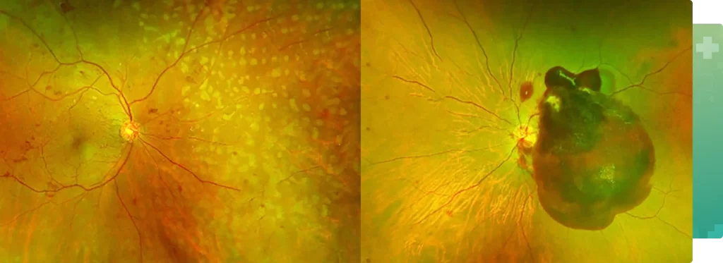
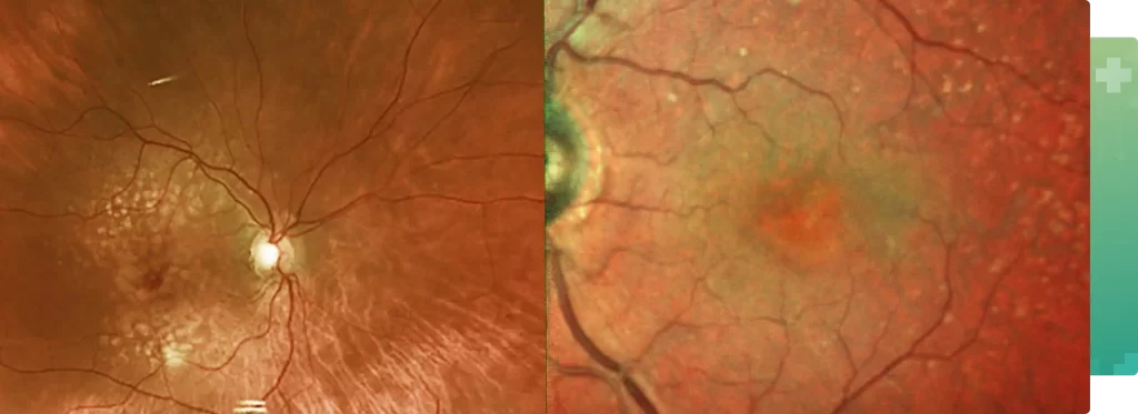
43-year-old gentleman visited to outpatient department with complain of diminution of in right eye for 1 week. On examination patient had severe meibomitis in left eye else anterior segment findings were within normal limits in both eyes. Patient was advised for dilatation and on fundus examination right eye revealed supero-temporal branch retinal vein occlusion with vitreous hemorrhage in right eye and left eye was within normal limits. He was advised for retinal imaging for both eyes with OPTOS and OCT for better understanding of the disease and explaining him the treatment required. There was no macular edema; with visible neovascularization noted along the superotemporal arcade. Patient was advised for superior sectoral PRP in right eye and asked for regular follow-up. By providing timely management with laser photocoagulation, we were able to prevent a massive vitreous hemorrhage and subsequent severe loss in vision. Patient is doing well currently and he is extremely happy with our treatment.
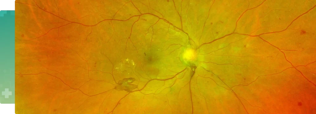
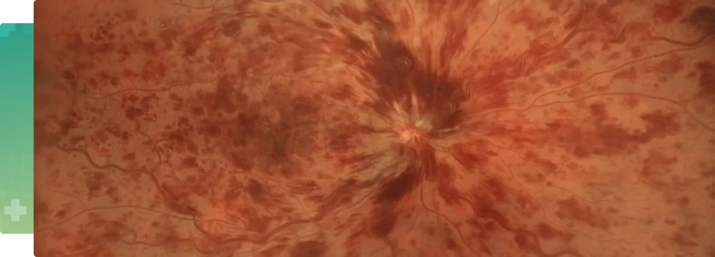
66-year-old lady visited to retina department in Narayana Nethralaya with complain of dimness of vision in right eye more than left eye for 8 months. She is known case of hypertension and diabetes mellitus for last 9 years. On examination anterior segment was within normal limits and advised for dilatation. Fundus examination in right eye revealed extensive hemorrhages and macular thickening in right eye and hemorrhage in the vitreous cavity in the left eye. Both the eyes findings were suggestive of advanced grade of diabetic retinopathy responsible for the poor vision in both eyes. She was advised retinal imaging with fluorescein angiography and optical coherence tomography for both eyes. FFA left eye shown neovascularization elsewhere in left eye with non-center involving diabetic edema in both the eyes. She was advised for retinal laser and intravitreal anti VEGF injection for left eye. Currently, patient improved visual acuity in left eye with stable vision in right eye. She is very happy and recovered her vision.
At Narayana Nethralaya, “Quality of Care” and “Patient Safety” is our priority. Concern for our patients’ well being is at the core of what we do, and what drives us. Five units of Narayana Nethralaya are NABH Accredited – the highest national recognition for quality in patient care and safety. Our Retina team help patients make an informed treatment choice on the type of treatment and surgery that is best suited for their lifestyle. We have an exclusive counseling team to address any doubts or questions that people may have about Retina treatment options and procedures.